
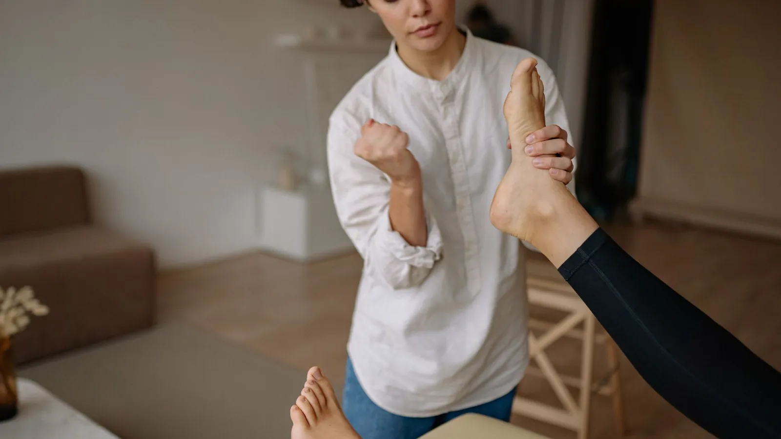
What it is
Plantar fasciitis is pain under the heel from the plantar fascia, the thick band of tissue that runs along the sole from the heel bone to the base of the toes. It is the most common cause of heel pain we see and one of the most common foot problems anywhere: roughly one person in ten has it at some point [2][11], and it accounts for around a million doctor visits a year in the United States alone [3].
Despite the name, the tissue is not inflamed. Samples taken from people with long-standing pain show a fascia that has degenerated and thickened under repeated load, with no inflammatory cells in it [5]. That changes the treatment. Rest and anti-inflammatories treat inflammation; this is a loading problem, and it responds to load applied in the right dose.
How it presents
- A sharp, stabbing pain under the heel with the first steps of the morning, easing within ten minutes or so of walking
- The same stab after sitting for a while, and a duller ache by the end of a long day on your feet
- Tenderness when the inside front edge of the heel bone is pressed
- Worse barefoot on hard floors, better in supportive shoes
- Usually one foot, sometimes both
The mornings are worst for a mechanical reason. Overnight the foot rests pointed down and the fascia sits slightly shortened, so the repair that happens during sleep happens at that shortened length. The first steps load it suddenly and stretch it out, hence the stab, and hence why it eases as the tissue warms and lengthens. The same thing happens after an hour at a desk.
Who gets it and why
It is a loading problem far more often than a structural one. Tissue that has adapted to a certain amount of load usually copes; it is the rate of change that causes trouble. Almost every case has a change behind it: more walking or running than before, a new job on hard floors, a switch to flat shoes, weight gained over a short period.
The largest case-control study of risk factors found three things that stood on their own: a stiff ankle, body weight, and a working day spent standing [4]. A tight calf that limits how far the ankle bends was by far the strongest, which is why the calf is treated as hard as the heel.
Risk factors
- Reduced ankle bend (dorsiflexion): people with no movement past neutral had over twenty times the risk of those with a free ankle [4]
- A body mass index above 30, with about five times the risk [4]
- Spending most of the working day on your feet, about three and a half times the risk [4]
- A recent increase in walking or running volume, or a change of footwear
- Age 40 to 60, when it peaks, though runners get it at any age and it makes up about a tenth of running injuries [2]
How it is diagnosed
Plantar fasciitis is a clinical diagnosis: the story of first-step pain and tenderness at the front of the heel bone is enough, and imaging is unnecessary in a typical case [2]. The assessment is mostly about ruling out the conditions that mimic it and about measuring the things that decide the plan, such as how far the ankle bends and how strong the calf is.
Heel spurs deserve a word, because many people arrive with an X-ray showing one and a belief that a bony spike is stabbing their foot. A spur is a sign of long-term traction on the heel bone, not a knife. In the largest long-term follow-up of plantar fasciitis, having a spur made no difference at all to how people did over the following years [11]. Removing it is rarely the answer.
When imaging earns its place
Imaging earns its place when the picture is atypical, when there is a suspicion of a stress fracture or nerve entrapment, or when a proper course of treatment has not worked. Ultrasound can confirm a thickened fascia; it does not change the first three months of treatment.
What the evidence says
Treatment by treatment, with the studies behind each verdict numbered to the sources at the end of the page.
Plantar fascia-specific stretching
Core treatmentPulling the toes back to stretch the fascia itself, ten holds of ten seconds, three times a day and always before the first steps of the morning. In people who had already had pain for ten months it beat calf stretching alone at eight weeks [7]; two years on, 92% were satisfied and 94% had less pain [7].
Progressive heel-raise strengthening
Core treatmentSlow, heavy heel raises with a rolled towel under the toes, every other day, adding load over weeks. In a trial of 48 people this produced clearly better function at three months than stretching; by six and twelve months both groups had improved similarly [6]. Loading gets you there faster and leaves a tissue that tolerates more.
Load management and footwear
Core treatmentRelative rest, not complete rest: fewer of the hours that flare it, supportive shoes indoors, cushioning at work if you stand all day. The guideline treats education about load and footwear as part of first-line care [1]. This is what stops you re-injuring it faster than it repairs.
Insoles and foot orthoses
Helps alongsideModerate-quality evidence of a small reduction in pain over the medium term, and off-the-shelf insoles did as well as custom-made ones in every comparison [8]. Useful support while the loading work does the real job; not a fix on their own.
Taping and hands-on treatment
Helps alongsideBoth are recommended in the guideline for short-term relief [1]. We use them in the early weeks to make walking tolerable, which is what lets you do the exercises.
Night splints
Helps alongsideA splint that holds the ankle near neutral overnight, so the tissue does not repair short, is supported in the guideline for pain that has lasted more than a few months [1]. Worth it for stubborn morning pain; most people do not need one.
Shockwave therapy
Second lineBetter than sham treatment in pooled trials [12] and ranked among the more effective options in a network analysis of 31 trials [9]. A reasonable next step when a proper course of loading has not worked, not a first step.
Corticosteroid injection
Use with cautionThe Cochrane review found low-quality evidence of a slight reduction in pain for up to a month, and none after that [10]. Repeated injections into a tissue that is not inflamed carry a risk of rupturing the fascia [5]. We do not suggest it as a first step; it has a place when pain is severe enough to block rehabilitation.
Ice, anti-inflammatories, massage guns, rolling
Helps alongsideIce after activity and a short course of anti-inflammatories are reasonable for comfort [2]. So is rolling the sole, within reason. None of them changes the tissue, so none of them is the plan.
Complete rest
AvoidWeeks off your feet make the calf weaker and the fascia less tolerant, and the pain returns the day you go back to normal life. Reduce the load; do not remove it.
Surgery
Second lineAbout 5% of cases end up in surgery, with inconsistent results [2]. It is a last resort after a year or more of properly delivered treatment, not an alternative to it.
Read together, the evidence is humbling. A 2019 network analysis of 31 trials could not show that any common treatment clearly beats the rest, though exercise was the one associated with long-term benefit [9]. Our reading of it: the early weeks are about comfort, with stretching, taping, footwear and an insole if it helps, and the months are about loading. The people who stay stuck are usually the ones who stretched, felt a bit better, and never progressed to the strengthening.
How we treat it
At The Rym Space, Bagalur
- 1
Assessment
Where it hurts and when, what changed before it started, your footwear and your day. We measure ankle bend, test calf strength and rule out the look-alikes: the Achilles, a nerve, the spine.
- 2
A plan you start that day
The morning routine, done sitting on the edge of the bed before the first step: a fascia stretch and a calf stretch. Footwear changes, taping if walking hurts, and why the spur on your X-ray is not the problem.
- 3
Week 1: settle the heel
3 to 5 sessions close together. Hands-on treatment and taping to take the sting out of walking, the stretches made a habit, footwear sorted, and the week's load trimmed to what the foot tolerates.
- 4
Week 2 onwards: build tolerance
Heel raises with a towel under the toes every other day, daily stretching, and hands-on treatment while the programme takes hold. Sessions space out as the home programme carries the work.
- 5
Weeks 6 to 12: load up and return
Single-leg heel raises, then added weight. Walking and running rebuilt on a graded plan, not on how it feels that morning.
- 6
Discharge, or the next tier
Done when first steps have been pain free for two weeks, you can do loaded single-leg heel raises, and you are back to what you came in for. Not on track by 8 to 12 weeks: we say so, and talk through a night splint, shockwave or, rarely, an injection.
What to expect, and when
Ranges, not promises. Where a figure comes from a study, the study is numbered.
- First 1 to 2 weeks
- The morning stab eases once the pre-step stretch is a habit. First-step pain responds earliest to fascia stretching [7].
- By 3 months
- In the strengthening trial, people doing loaded heel raises had clearly better function by now than those only stretching [6]. Most people on a consistent programme are largely pain free in daily life by three to six months.
- By 12 months
- About three quarters of cases have resolved within a year, with or without treatment [2]. The one in ten that persist [7] need the next tier, not more of the same.
- If it is left
- In hospital-referred cases, where the pain had already persisted, four in five still had it at one year and half at five years [11]. Early treatment is the difference.
The two long-term figures count different people: primary care, where most cases settle within a year, and specialist clinics, which see the ones that did not. Women and people with pain in both heels did worse in that study [11]. Do not wait months before starting a proper programme.
When to be seen sooner
Signs that this is something else, or that it needs looking at now rather than after a few weeks of self-care.
- Pain at the back of the heel rather than underneath, which points to the Achilles insertion
- Numbness, tingling or burning spreading into the sole, which suggests a nerve entrapment
- A heel that is painful to squeeze from the sides, especially after a sharp increase in running, until a stress fracture is ruled out
- A sudden tear or pop under the arch with bruising, which may be a fascia rupture
- Both heels becoming painful at the same time without a change in activity, particularly in someone under 40, which can be a sign of inflammatory arthritis
- Heel pain in a child or teenager, which is usually a growth plate problem and is managed differently
- Pain at night unrelated to activity, or feeling generally unwell with it
Common questions
- Do I need an X-ray or a scan?
- Usually not. The diagnosis is made from your story and the examination, and imaging does not change the first three months of treatment [2]. We will ask for it if something does not fit, or if a proper course of treatment has not worked.
- Should I stop walking or running?
- Reduce it, do not stop. Cut the volume to what the foot tolerates without a next-morning flare, keep supportive shoes on, and rebuild the distance on a plan once the loading programme is under way.
- Will an injection fix it?
- It may take the edge off for up to a month and the evidence does not show benefit beyond that [10]. Because the tissue is not inflamed, repeated injections risk rupturing it [5]. We keep it for pain that is stopping you doing the rehabilitation.
- Should I buy custom insoles?
- An off-the-shelf insole did as well as a custom one in every trial that compared them [8]. Buy a cheap supportive pair, wear them in your indoor shoes too, and spend the money you saved on decent footwear.
- How long until I can run again?
- It depends on how long you have had it and how far the calf has to come. As a rule of thumb, two weeks of pain-free first steps and a strong single-leg heel raise are the markers we want before running restarts, typically six to twelve weeks in.
Further reading
Sources
Guidelines, systematic reviews and trials only. Each number in the text links to the entry here.
- 1.Koc TA Jr, Bise CG, Neville C, Carreira D, Martin RL, McDonough CM. Heel Pain - Plantar Fasciitis: Revision 2023. Clinical practice guideline. J Orthop Sports Phys Ther. 2023;53(12):CPG1-CPG39. pubmed.ncbi.nlm.nih.gov ↗
- 2.Buchanan BK, Sina RE, Kushner D. Plantar Fasciitis. In: StatPearls. Treasure Island (FL): StatPearls Publishing; updated 7 January 2024. ncbi.nlm.nih.gov ↗
- 3.Riddle DL, Schappert SM. Volume of ambulatory care visits and patterns of care for patients diagnosed with plantar fasciitis: a national study of medical doctors. Foot Ankle Int. 2004;25(5):303-310. pubmed.ncbi.nlm.nih.gov ↗
- 4.Riddle DL, Pulisic M, Pidcoe P, Johnson RE. Risk factors for plantar fasciitis: a matched case-control study. J Bone Joint Surg Am. 2003;85(5):872-877. pubmed.ncbi.nlm.nih.gov ↗
- 5.Lemont H, Ammirati KM, Usen N. Plantar fasciitis: a degenerative process (fasciosis) without inflammation. J Am Podiatr Med Assoc. 2003;93(3):234-237. pubmed.ncbi.nlm.nih.gov ↗
- 6.Rathleff MS, Mølgaard CM, Fredberg U, et al. High-load strength training improves outcome in patients with plantar fasciitis: a randomized controlled trial with 12-month follow-up. Scand J Med Sci Sports. 2015;25(3):e292-e300. pubmed.ncbi.nlm.nih.gov ↗
- 7.DiGiovanni BF, Nawoczenski DA, Lintal ME, et al. Tissue-specific plantar fascia-stretching exercise enhances outcomes in patients with chronic heel pain: a prospective, randomized study. J Bone Joint Surg Am. 2003;85(7):1270-1277. Two-year follow-up: DiGiovanni BF, et al. J Bone Joint Surg Am. 2006;88(8):1775-1781. pubmed.ncbi.nlm.nih.gov ↗
- 8.Whittaker GA, Munteanu SE, Menz HB, Tan JM, Rabusin CL, Landorf KB. Foot orthoses for plantar heel pain: a systematic review and meta-analysis. Br J Sports Med. 2018;52(5):322-328. pubmed.ncbi.nlm.nih.gov ↗
- 9.Babatunde OO, Legha A, Littlewood C, et al. Comparative effectiveness of treatment options for plantar heel pain: a systematic review with network meta-analysis. Br J Sports Med. 2019;53(3):182-194. pubmed.ncbi.nlm.nih.gov ↗
- 10.David JA, Sankarapandian V, Christopher PR, Chatterjee A, Macaden AS. Injected corticosteroids for treating plantar heel pain in adults. Cochrane Database Syst Rev. 2017;6:CD009348. pubmed.ncbi.nlm.nih.gov ↗
- 11.Hansen L, Krogh TP, Ellingsen T, Bolvig L, Fredberg U. Long-term prognosis of plantar fasciitis: a 5- to 15-year follow-up study of 174 patients with ultrasound examination. Orthop J Sports Med. 2018;6(3):2325967118757983. pubmed.ncbi.nlm.nih.gov ↗
- 12.Tung WS, Daher M, Covarrubias O, Herber A, Gianakos AL. Extracorporeal shock wave therapy shows comparative results with other modalities for the management of plantar fasciitis: a systematic review and meta-analysis. Foot Ankle Surg. 2025;31(4):283-290. pubmed.ncbi.nlm.nih.gov ↗
Book an appointment
Start with a plantar fasciitis assessment
Your first appointment is a full assessment: history, hands on examination and movement testing, followed by an explanation of what we found and the plan to fix it. You leave with something to do that day, not just a follow up booking.
Monday to Saturday: 8:00 am to 1:00 pm, 4:00 pm to 8:00 pmSunday: By appointment